
Ever wonder how a simple eye exam can help keep your vision safe? Early checks can catch tiny changes like small yellow spots or a thinner central retina (macula) before you notice any problems. By getting your eyes examined on a regular basis, you and your doctor can act quickly to slow down vision loss. In this article, you'll learn how spotting these early signs lets you take some easy steps to protect clear sight for years to come.
Crucial Role of Early Detection in Macular Degeneration
Age-related macular degeneration (AMD) affects the center of your retina. It can make reading, driving, or recognizing faces harder by blurring your central vision. AMD starts quietly, with small changes like a thinning macula and tiny yellow spots called drusen, even when regular eye tests look normal. Regular, detailed eye exams can catch these early signs before more damage occurs.
Finding drusen and other subtle markers early means you and your doctor can step in before vision loss becomes noticeable. Today, eye care professionals use tools like optical coherence tomography (an imaging test that shows cross-sectional pictures of your retina) to spot even minimal changes. This early detection opens the door for timely actions such as nutritional tweaks and lifestyle changes.
Studies show that treatments like AREDS2 vitamins, along with habits like quitting smoking and eating more leafy greens, can slow AMD’s progression by about 25%. Keeping up with annual or more frequent check-ups ensures that any new changes are managed quickly, helping protect your vision for the long run.
Identifying Risk Factors to Enhance Early Detection of Macular Degeneration
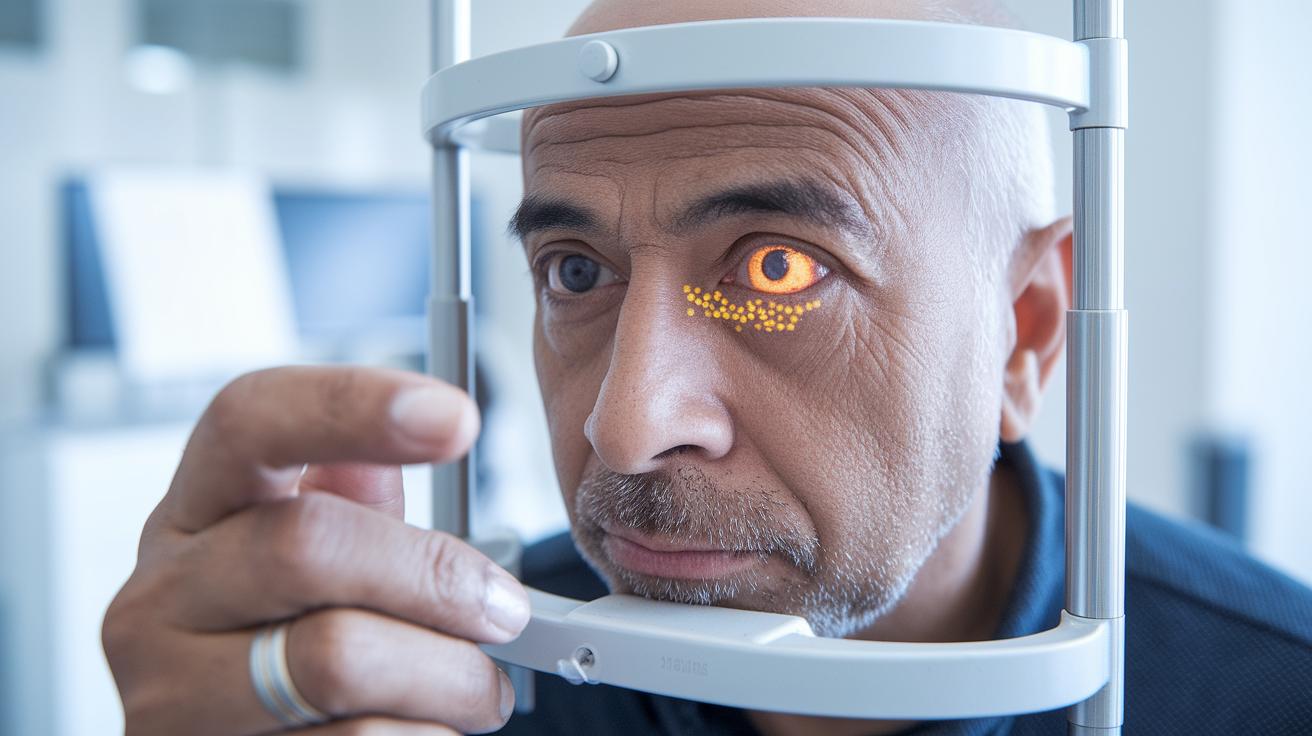
Knowing what can put you at risk for macular degeneration helps you catch its early signs. Research shows that once you hit 50, your risk goes up, and factors like your lifestyle and genes can add to that risk. For example, changes in genes like CFH and ARMS2 may increase your chance of developing this condition. Doctors can spot early warning signals, such as small yellow deposits on the retina called drusen, by checking for changes related to aging. With genetic tests and careful age assessments, they can create a plan just for you. This plan might include early advice on nutrition and lifestyle changes, along with regular eye exams to catch any issues before they worsen.
Here are common risk factors to consider:
- Being older than 50
- Smoking, past or present
- High blood pressure
- A family history of macular degeneration (AMD)
- Eating a diet low in antioxidants
By understanding these risks, you and your doctor can take proactive steps to catch macular degeneration early and protect your vision.
Advanced Diagnostic Imaging Techniques for Early Macular Degeneration
Optical Coherence Tomography (OCT) is a key tool in spotting early macular degeneration. It shows a detailed, layered view of your retina, helping doctors see tiny deposits (drusen), areas where the retinal pigment epithelium is thinning, and even small pockets of fluid that might be missed during a routine exam. Color fundus photography also plays a big part by capturing a wide view of the retina, letting clinicians count and track these deposits. Using OCT and fundus photography together means fewer early signs slip by, so you can get timely advice on slowing the condition with simple lifestyle tweaks.
Other imaging methods add even more value when used together. Fundus autofluorescence maps out spots where the retinal cells may be under stress. Meanwhile, OCT angiography is a growing, noninvasive way to see the tiny blood vessels (called the choriocapillaris) that support the retina. This technique detects small changes in blood flow that might hint at a shift to a more active stage of the disease. By comparing different types of images, doctors can more accurately spot subtle signs of degeneration and tailor treatment for each person. With technology constantly improving, these techniques are vital for giving every patient the best early care possible.
| Imaging Technique | Detection Purpose | Key Strength |
|---|---|---|
| OCT | Drusen, RPE thinning, fluid | Layered, cross-sectional view |
| Fundus Photography | Drusen count/distribution | Wide, colorful retinal image |
| Autofluorescence | RPE health | Highlights stressed retinal cells |
| OCT Angiography | Choriocapillaris flow | Noninvasive view of small vessels |
Symptom Awareness and Progressive Vision Monitoring in Macular Degeneration

Watching your vision closely can help you catch early signs of macular degeneration. Even small changes, like a subtle blur in the center of your sight, might be the first hint that something isn't right. These slight shifts can make everyday tasks, like reading a menu or recognizing a familiar face, more challenging. Noticing these changes early means you can mention them at your next eye exam, which helps catch any progression before it worsens.
Some people notice that lines seem wavy or that small areas in their vision appear dark or even blank. These changes might start in one eye before affecting the other. For example, you might see a tiny dark spot on your Amsler grid (a simple tool to test your central vision) during a quick check. Spotting these early signs opens the door to timely treatment and preventive steps.
Regular self-checks are a practical way to keep an eye on your vision. Using an Amsler grid at home makes it easier to spot any new distortions like cloudy or spotted images. If you notice any changes, contact your eye care professional right away. Early intervention can greatly increase your chances of preserving clear, central vision.
Regular Screenings and Clinical Protocols for Early Macular Degeneration Detection
If you're 50 or older, it's a smart move to schedule an annual eye exam. When you arrive, you'll fill out a quick health update, then go through a few short tests before the main eye check. The clinic explains every step so you always know what to expect. One patient said, "I felt reassured when my optometrist walked me through each part of the exam."
During your screening, your doctor uses a few simple tools one after the other. First, they check how clearly you can see at a distance. Next, they perform a fundus exam to take a close look at your retina (the back part of your eye). You'll also be asked if you’ve noticed any changes in your vision, even small ones. One patient shared, "My doctor explained each test clearly, which made me feel confident about catching early signs."
Today’s clinical steps follow proven guidelines like the AREDS classification (a system that helps rate eye health) to decide on follow-up visits and treatment options. With new telemedicine options, you might even get part of your checkup reviewed remotely. This lowers wait times and makes keeping an eye on your vision easier.
Preventative Vision Care and Lifestyle Interventions in Early Macular Degeneration

Research shows that AREDS2 supplements may lower the risk of worsening macular degeneration by about 25%. These supplements include 500 mg of vitamin C, 400 IU of vitamin E, 10 mg of lutein, and 2 mg of zeaxanthin. Studies back these vitamins as a strong defense for your eyes by helping reduce oxidative stress in the retina. One patient said, "My eye doctor recommended these vitamins during my check-up, and it made me feel more secure about keeping my vision healthy."
Your daily habits also make a big difference in eye health. Stopping smoking is one proven way to lower the chance of serious vision loss. Eating plenty of leafy greens and omega-3 fatty acids (healthy fats found in foods like fish) helps support the retina. Regular exercise and protecting your eyes from UV rays also reduce eye stress. Simple changes like choosing water over sugary drinks or taking a brisk 10-minute walk every day can add up to stronger, healthier vision over time.
Researchers are still studying new ways to help with early signs of macular changes, such as using higher doses of statin medications. While these methods are promising, they are still under investigation. By mixing new ideas with proven lifestyle habits, you can work towards keeping your vision clear and strong for everyday activities.
Innovations and Emerging Technologies in Early Detection of Macular Degeneration
New imaging tools are changing how doctors catch macular degeneration early. One standout tool, adaptive optics scanning laser ophthalmoscopy, lets clinicians see eye cells up close, giving them a clear view of your retina's condition. Smart computer programs (AI), meanwhile, check fundus photos and OCT scans to spot small changes that might slip by otherwise. OCT angiography adds another layer by mapping tiny blood vessel changes without needing dye. Just picture a machine that takes detailed pictures of tiny cells, finding signs long before your vision feels different.
Portable retinal scanners and video-based eye care (teleophthalmology) are also making early screenings more accessible. These advances bring diagnostic power to remote and underserved areas so more people can get speedy evaluations. Researchers are now studying new markers (biomarkers) that may pick up early stress on the retinal pigment epithelium (RPE), a key part of your eye, even before symptoms appear. Imagine a compact, hand-held scanner that gives quick feedback on your eye health, offering hope for even earlier treatment.
Final Words
In the action of early detection of macular degeneration, we saw how comprehensive eye screening, diagnostic eye exams, and risk factor evaluation work together to catch subtle retinal changes early.
Simple self-checks and using tools like the Amsler grid can signal the need for timely professional advice.
Combining these insights with smart supplement choices and lifestyle habits can help protect your vision. Small, informed steps can make a real difference every day.
FAQ
I cured my macular degeneration?
The claim of curing macular degeneration reflects personal experience. Research shows that treatments aim to slow progression, not completely cure AMD, making early detection crucial.
What vision looks like with macular degeneration?
Descriptions of vision with macular degeneration often include blurred central detail, wavy lines, and dark spots. These signs indicate that changes in the macula may be impairing clarity.
How to prevent macular degeneration?
Preventing macular degeneration involves healthy lifestyle choices like avoiding smoking, eating nutrient-rich foods, and using supplements as recommended. Regular eye exams also help catch early changes.
What is the main cause of macular degeneration?
The main cause of macular degeneration is the gradual aging process, where changes such as drusen deposits affect the macula. Genetic factors and environmental exposures also play a role.
What is dry macular degeneration?
Dry macular degeneration is the slower, more common form of AMD marked by drusen deposits and thinning of the retinal tissue. It is often managed with nutritional support and lifestyle adjustments.
What is the best treatment for macular degeneration?
The best treatment for macular degeneration focuses on slowing progression through AREDS2 supplements, lifestyle improvements, and, in some cases, anti-VEGF therapies for wet AMD. Personalized care is key.
What does macular degeneration surgery involve?
Macular degeneration surgery is rarely used and is typically reserved for advanced wet AMD cases. It addresses complications like bleeding or scar tissue and is individually evaluated by specialists.
Can macular degeneration be detected early?
Early detection of macular degeneration is achievable through comprehensive eye exams using advanced imaging like OCT. Identifying changes before severe symptoms appear enables prompt management.
What are the first signs of macular degeneration?
The first signs of macular degeneration include subtle blurry spots in central vision and slight distortions, like wavy lines. Recognizing these symptoms early can lead to timely evaluation and care.
What is the average age for getting macular degeneration?
Macular degeneration typically appears in people aged 50 and older. Aging increases the risk of retinal changes, making regular screening important as you enter midlife.
Can macular degeneration be stopped if caught early?
Detecting macular degeneration early allows for interventions that slow its progression. Although early measures can prevent rapid decline, the condition is usually managed rather than completely halted.